The pioneering technique that avoids opening the abdomen to save a vital vein in children
The Vall d’Hebron Hospital operates on the obstructed portal vein, which can go unnoticed for years, with minimally invasive surgery
-
CAT -
CAST
BarcelonaMaría José Correa was 12 years old when she began vomiting large amounts of blood. She had never experienced any serious health problems, and her family was terrified. Doctors in her native Vitoria identified that she was suffering from a digestive hemorrhage caused by a clot in the portal vein, which is responsible for transporting blood from the intestine and spleen to the liver. The girl had to be urgently transferred to the Vall d'Hebron Hospital, where she underwent a pioneering, minimally invasive operation that was a complete success. In fact, by the next day, she was already able to lead a completely normal life, and today she has no sequelae or complications.
When the portal vein becomes obstructed, blood cannot circulate normally and seeks alternative paths, which causes an increase in blood pressure in the vein, an enlargement of the spleen, a decrease in platelets, and the appearance of varices in the esophagus and stomach that can end up causing serious hemorrhages. Until now, one of the main alternatives for restoring blood circulation was a complex surgical intervention, known as a Rex shunt, which consists of creating a new path for blood to reach the liver.
Pediatric surgeon José Andrés Molino has explained that these operations require opening the abdomen and, in some cases, extracting a vein from another part of the patient's body to build the new connection. These are interventions lasting several hours, with a significant risk of bleeding, which usually entail the patient spending a few days in the intensive care unit (ICU) and at least a week hospitalized. In contrast, his new procedure usually lasts between two and three hours and recovery takes less than 24 hours.
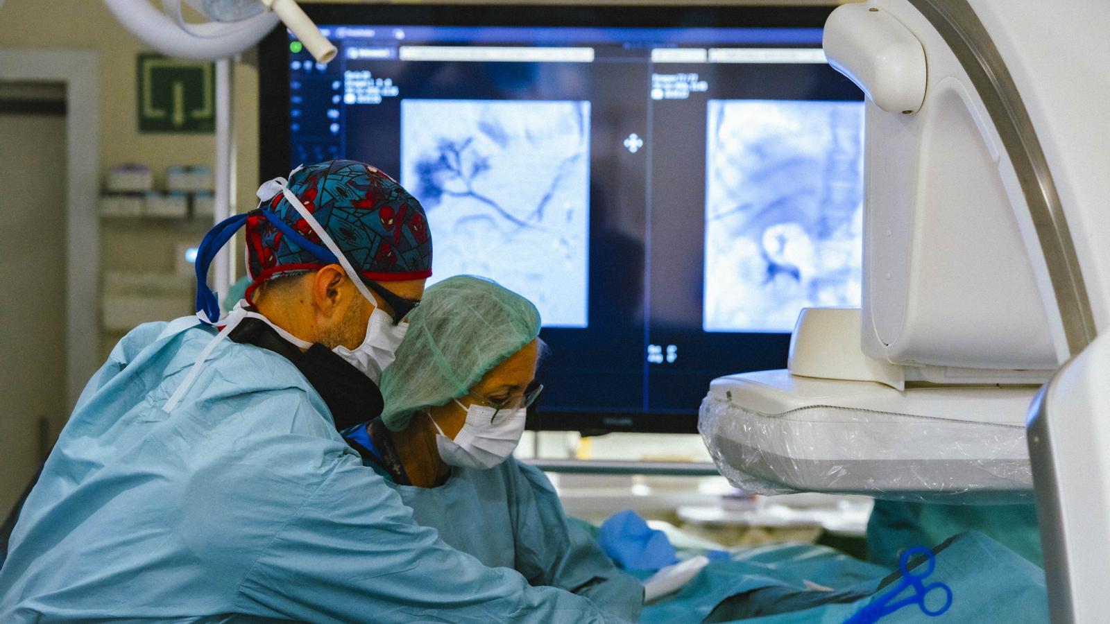
This pioneering technique is performed with two small punctures—one through the spleen and another through the liver—and allows for the restoration of blood circulation and the prevention of serious complications, such as digestive hemorrhages. With the help of radiological imaging, specialists introduce catheters until they reach the obstructed vein. Once they gain access, they attempt to reopen it with small balloons and place a metallic prosthesis (stent) to keep the blood flow open.
Since 2018, the Vall d'Hebron Hospital has managed to reopen the portal vein in 12 of the 24 children treated with this technique. Of these 12 patients, 10 currently maintain an open vein. However, the technique is not effective in all cases, and the choice of treatment depends on the characteristics of each patient and their vascular anatomy. Mercedes Pérez, head of the interventional vascular radiology section at the hospital, has highlighted that the procedure allows one in two children to avoid complex surgery.
Specific protocol for newborns
Unlike adults, in whom this condition is associated with cirrhosis, in children it is often caused by portal vein thrombosis. Risk factors include prematurity, coagulation disorders, and the placement of catheters through the umbilical cord during the first days of life. However, in more than half of the affected children, a clear cause of the obstruction is not identified, not even after an exhaustive medical investigation.
This extrahepatic obstruction can go unnoticed for years and manifest itself for the first time in pre-adolescence or adolescence, usually between 10 and 15 years of age. The average age of the treated children is 8 years. For this reason, the Barcelona hospital now has a follow-up protocol with ultrasounds for babies who have been fitted with an umbilical catheter to detect possible thrombosis before it causes complications in the future.
The head of the pediatric hepatology and liver transplant section at Vall d'Hebron, Jesús Quintero, explained that "only three or four centers in the world perform this type of intervention" and that, when they presented the first results of the new technique in international forums of specialists, some professionals were incredulous at the possibility of resolving with such a minimally invasive procedure a disease that until then required highly complex surgeries.
The Barcelona hospital receives patients from other autonomous communities, as it is a rare pathology that requires highly specialized teams. Doctors have not been able to specify its incidence in Catalonia or Spain because there are no sufficiently complete records. Currently, however, Vall d'Hebron is collaborating with a hospital in Madrid so that it can also perform the procedure.
